
Over the past 16 years, Kim has worked as a general duty, senior, and charge sonographer. She began her career as a new graduate in Brandon before moving to St. Boniface Hospital in Winnipeg, where she has spent the majority of her career.
An Extension of the Radiologist
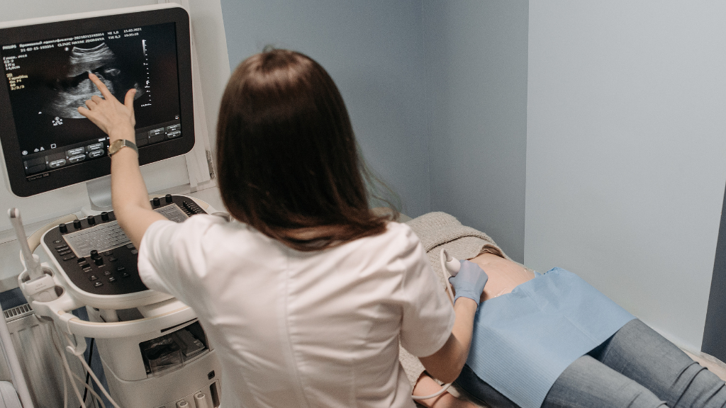
Sonographers are on the frontline of imaging, with radiologists relying on their critical thinking and attention to detail to guide diagnosis and patient care. In many countries, radiologists perform ultrasounds themselves, whereas in Canada, sonographers act as an extension of the radiologist—a unique partnership that sets the profession apart from other imaging disciplines.
“We connect the dots between the referring physician and the radiologist,” Kim explains. “By uncovering key findings and building relationships with patients, we help ensure their stories and symptoms translate into meaningful answers and care.”
St. Boniface Hospital is one of only two sites in Manitoba where sonographers work directly with sonologists. These experts built the foundation for the province’s standard of care and helped shape ultrasound practice across Canada, even laying the groundwork for the local training program. Kim describes their mentorship as transformative: the sonologists challenge sonographers to think critically and uphold the highest standards of quality in their work.
Over the past five years, the field has begun to shift as many of these experts have reached—or are nearing—retirement. Now, their former students who have become experienced sonographers themselves are stepping into the role of expert scanners, determined to carry forward the high standards instilled in them.
“We’re driven to keep ultrasound an integral and respected component of medical imaging because we know the value we provide to patients and to the health-care system,” Kim says.

It was at St. Boniface that Kim found the space to truly grow—benefiting from the mentorship of skilled and experienced sonographers and sonologists, the specialized radiologists who work exclusively in ultrasound. These experts were deeply committed to exceptional patient care and to using ultrasound in innovative ways. Working alongside them, Kim saw how sonographers could drive meaningful change within their field. She witnessed teams collaborating with specialists to pioneer new processes for detecting congenital heart defects and saw firsthand how innovation could directly transform patient care.
Though initially drawn to ultrasound by a general interest in medicine, Kim quickly realized the profession sparked her inner drive for constant growth and development, while allowing her to play a vital role in patient care. A graduate of the University of Winnipeg and the Health Sciences Centre School of Diagnostic Ultrasound, Kim holds a Bachelor of Science in Biology and a Diploma in Diagnostic Medical Sonography.
“I chose this profession because I want to help people,” she says. “Ultrasound allows me to uncover what’s hidden beneath the surface and help patients in meaningful ways.”
More than just ‘taking pictures’
Ultrasound is safe, accessible, and cost-effective, often making it the first test ordered by clinicians—this makes sonographers like Kim among the first healthcare professionals a patient meets on their diagnostic journey. Ultrasound plays a critical role in uncovering life-changing findings, from detecting cancer to identifying congenital defects in babies.
A typical day for Kim involves up to 11 scheduled outpatient exams or a focus on our inpatients and those in the Emergency Department. Each case varies in complexity and urgency, and unexpected findings often require extra time, focus, and coordination with radiologists or referring physicians.
“An exam involves far more than just taking pictures,” she explains. “I spend 30–60 minutes with each patient—listening to their concerns, connecting their story to what I see on the screen, and carefully evaluating each organ while adjusting the settings to get the clearest possible images. I then share a detailed summary of my findings with the radiologist. Every exam is unique, and we often adapt in real time to ensure a complete exam—helping to guide a patient’s next steps in care.”
A key distinguishing feature of ultrasound is its ability to evaluate anatomy through dynamic movement. Sonographers can pinpoint specific areas to apply pressure, assess for tenderness, observe tendon or ligament motion, and even evaluate the natural sliding of organs. No other imaging modality offers this interactive assessment, and the information it provides is often critical in determining the need for surgical intervention.
Advocating for Women’s Health: Advancing Endometriosis Care
It’s estimated that as many as 50% of women with chronic pelvic pain have endometriosis. The disease causes severe pain, fertility challenges, and reduced quality of life, yet diagnosis often takes 7–11 years on average.
Throughout her career, Kim has witnessed continuous advances in patient care across the province. As a young sonographer, she was inspired by colleagues who proved that sonographers could be catalysts for meaningful change. Now, with years of experience, mentorship, and tens of thousands of scans behind her, Kim is working to drive progress in a long-underserved area of women’s health.
“After countless experiences—both professionally and personally—with women suffering from debilitating pelvic pain, I became passionate about how we can better support these women through ultrasound.”
Many of these women undergo repeated physician visits, multiple pelvic ultrasounds, CT scans, and lengthy emergency department stays, only to be told that no abnormality was found. It’s estimated that as many as 50% of women with chronic pelvic pain have endometriosis—a condition where uterine tissue implants on other organs in the pelvis, and sometimes even in the upper abdomen. The disease causes severe pain, fertility challenges, and reduced quality of life, yet diagnosis often takes 7–11 years on average.
Only recently have technological advances allowed for the detection of sub-centimeter lesions with remarkable clarity—sometimes even surpassing the resolution of other imaging modalities. However, this progress relies on pairing advanced technology with the skill, precision, and critical thinking that experienced sonographers bring to each exam. These advancements are the result of extensive international research and development, which have refined the specialized techniques required for accurate detection.
For the past 18 months, Kim has been collaborating with a team of sonographers, radiologists, and a gynecological expert in endometriosis to develop a new scanning protocol aimed at improving detection and diagnosis. The work has required extensive research, hands-on training, repeated case exposure, and close correlation with other imaging modalities.
“A lot of this work has taken place without dedicated time or formal support, within a system that still lacks a structured process for managing these patients,” Kim says. “But we’re beginning to see real progress. Knowledge is spreading, we’re gaining a clearer picture of how to refine our process, we are building confidence in our findings, and patients are starting to see tangible improvements in care.”
While much of this work is still in its early stages, Kim and her team aspire to share their knowledge and experience across the province.
“I can’t fix the entire system, but I can still make a direct and meaningful difference for every patient I scan.”
“We aim to engage the skills and expertise of our fellow sonographers, enabling them to play an integral role in a more streamlined and effective process—one that reduces diagnostic delays and ultimately improves patient outcomes,” she says.
The potential impact on women’s health is profound, with wider benefits for the health-care system—reducing repeat exams, lowering emergency department visits, and shortening diagnostic wait times.
“The greatest reward is seeing the immense gratitude patients have for the work we do. These are powerful moments, reminding me that while I can’t fix the entire system, I can still make a direct and meaningful difference for every patient I scan.”